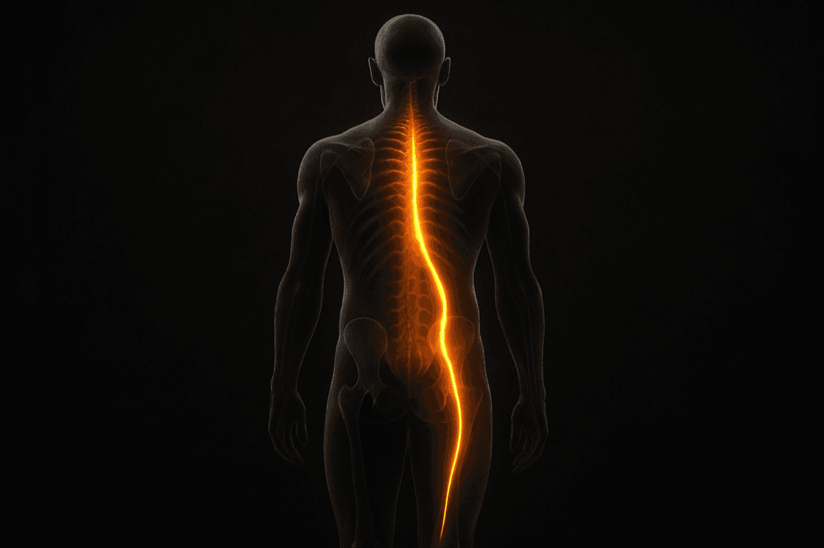
What Sciatica Is and Is Not
Sciatica is the term for pain, numbness, tingling, or weakness that follows the path of the sciatic nerve: from the lower back through the glute, down the back of the thigh, into the calf, and sometimes into the foot. It is one of the most common complaints bringing adults to medical care, and one of the most commonly misunderstood.
Sciatica is a symptom, not a diagnosis. The pain is the sciatic nerve expressing irritation or compression. The cause of that irritation or compression is what varies, and that cause is what determines whether treatment is effective.
The most commonly cited cause is lumbar disc herniation: a disc between vertebrae L4-L5 or L5-S1 bulging into the space where the sciatic nerve root exits the spinal canal. This is accurate as far as it goes. Disc herniation can compress a nerve root and produce true radicular pain. But it is not the only cause, it is not always the primary cause, and the presence of a disc herniation on imaging does not necessarily mean that herniation is producing the symptoms.
This is the first thing doctors sometimes miss: correlation between imaging findings and symptoms is surprisingly weak for lumbar disc herniations.
The Imaging Problem
A landmark study published in the New England Journal of Medicine examined MRI findings in adults with no back pain or sciatica whatsoever. Among people in their fifties, approximately forty percent had disc abnormalities visible on imaging, including bulges, protrusions, and herniations, that were completely asymptomatic. By their sixties, the percentage was higher.
This means that when a patient presents with sciatica and an MRI reveals a disc herniation, the clinician faces a critical question: is this herniation causing the sciatica, or is it an incidental finding in a patient who also has sciatica from a different cause?
The answer matters enormously because the treatments are different. A herniation causing nerve root compression may require medical management or, rarely, surgery. A piriformis muscle compressing the sciatic nerve in the glute, which produces identical symptoms, requires entirely different treatment. An anterior pelvic tilt driving chronic lumbar compression that is irritating the nerve without a discrete herniation requires structural corrective work.
The imaging reveals anatomy. It does not reveal mechanism. And mechanism is what treatment must address.
The Piriformis Problem
The piriformis is a small, deep muscle in the glute that runs from the sacrum to the top of the femur. The sciatic nerve passes directly beneath it, and in a significant percentage of the population, through it.
When the piriformis becomes hypertonic, meaning chronically contracted, it compresses the sciatic nerve. The symptoms are indistinguishable from disc-related sciatica: pain radiating from the glute into the back of the thigh and calf, numbness, tingling.
Piriformis syndrome is almost certainly underdiagnosed. It does not show up on MRI. The piriformis is not easily imaged in a way that reveals its state of contraction. A clinician looking at an MRI that shows a disc bulge in a patient with sciatica has a structural explanation that looks compelling, even when the piriformis is the actual culprit.
The functional test is straightforward: a patient with piriformis syndrome will have significant tenderness to deep pressure in the glute at the piriformis, and the pain will be provoked by hip external rotation under load. Disc-related sciatica is typically worsened by spinal loading, including sitting, sneezing, and coughing. Piriformis syndrome is typically worsened by the specific hip position that compresses the sciatic nerve at that point.
Why does the piriformis become hypertonic? Almost always because the glutes are inhibited.
Free - 60 seconds
Not sure which program you need?
Take the free posture quiz and get a personalized recommendation based on your pain and goals.
The Structural Chain Nobody Explains
The piriformis is a hip external rotator. In a well-functioning hip, the gluteus maximus and gluteus medius do the heavy work of external rotation, extension, and abduction. The piriformis is a secondary contributor.
When the glutes are inhibited, which happens when anterior pelvic tilt places them in a mechanically disadvantaged position, the piriformis compensates. It takes on load that should be distributed across the larger gluteal muscles. Under this chronic overload, it becomes hypertonic. It tightens. It compresses the sciatic nerve.
The fix for piriformis syndrome is not stretching the piriformis, though this can provide temporary relief. The fix is restoring glute function by correcting the anterior pelvic tilt that inhibited the glutes in the first place.
This is the structural chain that most clinical management of sciatica does not address: anterior pelvic tilt inhibits glutes, glute inhibition causes piriformis overload, piriformis overload compresses sciatic nerve, sciatic nerve compression produces sciatica symptoms.
Why Rest Makes It Worse
The standard medical advice for acute sciatica is rest. Do not bend, do not lift, avoid anything that provokes the pain. In the short term, this reduces the acute inflammatory response. In the medium term, it makes the underlying condition worse.
The sciatic nerve requires movement to maintain its capacity to glide through the tissues it passes through. Every joint, fascial layer, and muscle it passes from the lumbar spine to the foot is a potential source of restriction. The nerve is designed to glide freely through these structures as the leg moves. Extended rest allows adhesions to form, areas where the nerve becomes restricted in its movement through surrounding tissues. These adhesions produce symptoms that persist even after the original compression resolves.
Neural flossing, meaning specific movements that alternately tension and release the sciatic nerve through its full path, maintains this gliding capacity and is among the most effective interventions for sciatica that extends beyond the acute phase. They cannot be performed at rest.
The position that most reliably reduces acute sciatic nerve compression without loading the lumbar spine is static back: lying supine with hips and knees at ninety degrees. This position decompresses the posterior lumbar disc margins and facet joints while gently lengthening the hip flexors, addressing both disc-related and piriformis-related compression simultaneously.
The Structural Correction
For sciatica from any structural cause, whether disc-related, piriformis-related, or generalized lumbar compression, the corrective approach is identical because the structural driver is identical: anterior pelvic tilt creating lumbar compression and glute inhibition.
The sequence: static back to decompress the lumbar spine and begin restoring pelvic neutrality. Supine groin progressive to lengthen the hip flexors driving the tilt. Glute bridges to begin reactivating the inhibited posterior chain. Hip external rotation mobility work to restore the available range that takes load off the piriformis. Progressive return to full movement through neural flossing and functional movement patterns.
This approach works whether the compression is at the disc level or the piriformis level, because it addresses the structural state that produces both. It does not require a precise diagnosis of which mechanism is operative. It requires addressing the mechanism that underlies both.
Most people with chronic sciatica who implement this structural approach see meaningful symptom reduction within two to four weeks. Resolution of the structural driver takes eight to twelve weeks of consistent work. After that, the symptoms do not return unless the structural dysfunction returns, which ongoing maintenance prevents.
The question is not what is compressing the sciatic nerve. The question is why the body is in a structural state that allows that compression to happen. That is the question corrective exercise answers. For a structured approach to resolving sciatica from the structural root, the Sciatica Relief program covers the full sequence.
Mike Boshnack
Corrective Exercise Specialist · Posture Guy Mike
Mike Boshnack grew up skateboarding and surfing, trained MMA, and rode road bikes competitively. A shoulder injury put him on a path to discover corrective exercise. He has since helped thousands of people fix the structural patterns causing their pain, without surgery or passive treatments.
Keep Reading
Related Conditions
Free tool
See where your posture stands right now.
Upload a photo and get an instant AI-powered posture analysis with personalized recommendations. Free, no account required.
Try the Free Posture CheckTake the next step
Fix the structural root cause, not just the symptom.
Mike's programs apply this corrective method to your specific condition. No gym, no equipment. Just a floor and 15 minutes. Buy once, own forever.
Discussion
Discussion is a Pro member feature. Visit the community for more.